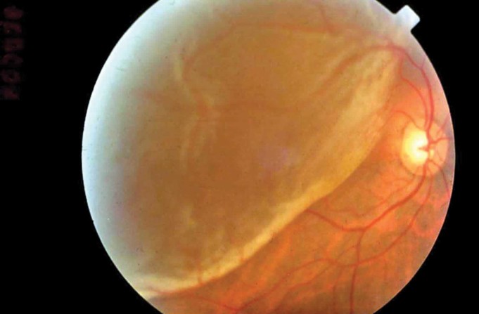
Retinal detachment is the process of separating the retina of the eye from the choroid. In a healthy eye, the retina is tightly attached to the choroid, from which it receives nutrition.
Without timely surgical treatment retinal detachment leads to blindness. Most often, it occurs with eyes damage and myopia, as well as with diabetic retinopathy, intraocular tumors, dystrophy of the retina, etc.
Causes of retinal detachment
To understand the reasons, you need to know the mechanism of the disease development.
It can be provoked by physical over strain or pressure on the surface of the retina that results in small defects via which liquid from the vitreous body moves into the space under the retina.
As a result, this fluid begins to separate the retina. The more fluid leaks out, the larger the detachment area will be. Basically, such a disease is observed only in one of the eyes, however, it negatively affects the other eye.
Therefore, it is important to examine both eyes together.
Risks factors and symptoms
Sometimes patients note that their vision improves slightly after sleep. This is due to the fact that with the horizontal position of the body the retina returns to its place.
When a person takes a vertical position, it departs from the choroid again and visual defects appear.
Symptoms of retinal detachment
- Blurring of the vision
- Flashes in the form of sparks and lightning
- Distortion of the letters.
Another symptom is loss of sight of in the individual retina sections. The risk factors leading to the retinal detachment are:
- Peripheral vitreoretinal retinal dystrophies.
- Retinal detachment in one of the eyes.
- Severe myopia.
- Eye injuries.
- Retinal detachment in close relatives.
Types of retinal detachment
Retinal detachment can be primary and secondary. Pathology is called primary when detachment is preceded by a rupture with subsequent leakage of fluid under the retina.
Secondary detachment develops as a complication of any pathological process, for example, due to the development of a neoplasm between the retina and choroid of the eye. The following types of retinal detachment can be distinguished:
- Rhematogenous (from Greek rhegma, rupture) arising from a rupture of the retina.
- Traction, arising due to tension of the retinal tissue from the vitreous body.
- Exudative, characterized by the penetration of serous fluid into the space under the retina. It arises due to increased vascular permeability and other hemodynamic disorders of the eye.
Retinal detachment treatment
Retinal detachment is a medical emergency requiring urgent surgical intervention to avoid total blindness. Surgery is the only option to treat this disease.
The earlier the operation is performed, the more chances regain vision and save the eye are. The task of surgical treatment for retinal detachment is to detect retinal rupture, close it and create a strong adhesion between the vascular and retinal membranes.
Depending on the type of retinal detachment, the surgeon will choose one of the specific methods or a combination of them:
- Local filling in the area of retinal rupture is carried out in cases when the retina is partially detached.
- Circular filling is used in more severe cases, when the retina has detached completely and there are multiple tears.
- Vitrectomy is a method in which a damaged vitreous is removed from the eye and one of the medications is injected: physiological saline, liquid silicone, perfluorocarbon compound in the form of a liquid or a special gas that presses the retina from the inside of the choroid.
- Laser coagulation is applied to limit the area of rupture and enhance thinned sections of the retina.
- Pneumatic retinopexy is a procedure when the retina specialist injects a gas bubble into your eye. This can help your retina to return back into her place.
Even after a successful therapy, the patient needs regular monitoring by an ophthalmologist.
It is performed 2 times a year with a thorough examination of all closed retinal tears. Such patients undergo regular courses of conservative treatment, including the injections of metabolic, retinoprotective, and vitamin preparations.
It is mandatory to exclude physical exertion and weight lifting for the rest of your life after the operation.
Retinal detachment prevention
If the patient has severe myopia with serious changes in the fundus or other pathology of the retina, it is recommended to visit an ophthalmologist once a year to examine the fundus.
It is better to control the regime of physical exertion and do not lift heavy objects. Actually, the same recommendations concern people with normal vision and no problems with the retina.
As we can see, retinal detachment is a dangerous disease that should be operated on immediately. You can do this abroad easily thanks to Booking Health.
With the help of the company you can find a suitable clinic with affordable prices for medical services and undergo retinal detachment treatment in the shortest possible time. Sooner you see a doctor, greater the chance of vision restoration is.


